

On May 31st, Dr. Pinnaro received the Excellence in Clinical Coaching award from the Graduate Medical Education office. This award is given to faculty who are distinguished as outstanding clinical teachers of resident and fellow physicians. Comments written about Dr. Pinnaro’s teaching include a reflection of her approach to hospital rounds where she is renowned for “probing each learner with thought-provoking questions to assess their understanding, empowering the learners to take ownership of the patients, and dropping clinical pearls about even the mundane patient.” Congratulations Dr. Pinnaro, and thank you for your outstanding clinical teaching efforts.
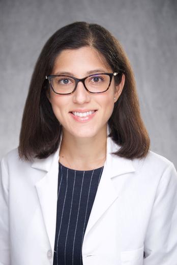
Drs. Curtis and Pinnaro Provide Invited Commentary on Pubertal Timing
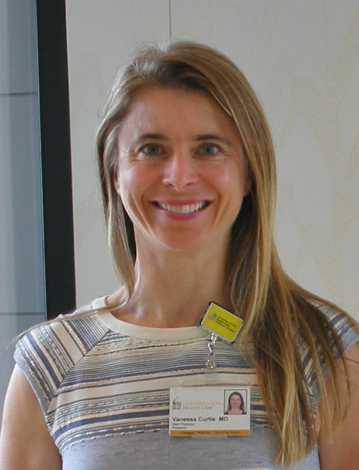
A variety of pathological processes can induce puberty earlier than otherwise would normally occur. When a child enters puberty, an important question is whether the timing is abnormally early. Pediatric endocrinologists are often the arbitrators of this question. Data defining the normal ages of puberty start are thus important. Data suggest that normal timing varies depending on a child’s genetics and environment. A recent publication in JAMA Network Open reports data collected from over 100,000 youth with Asian American, Native Hawaiian, and/or Pacific Islander heritage (link to article). To help interpret the findings, Drs. Vanessa Curtis and Catherina Pinnaro from our division were asked to provide their commentary on the article. Their commentary can be found here (link, which has link to the open source full text). They conclude that “[the] study […] emphasizes the necessity for precision and the pitfalls encountered when using race and ethnicity as a proxy for genetic background.”
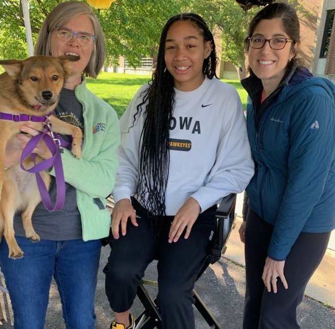
JDRF Diabetes Fundraising Walk
The JDRF is a philanthropic, nonprofit organization that raises funds to support research aimed at curing, preventing, and better treating type 1 diabetes. On Saturday, May 11, the JDRF held its annual fundraising walk in Cedar Rapids. Our Division, led by Drs. Pinnaro and Alexandrou, organized a team of walkers to help the cause. Walkers from our division on the team included Drs. Alexandrou and Pinnaro, nurse practitioner Alex, diabetes nurses Haylee and Sue, pharmacist Lisa, our administrator Teresa, and research coordinator Emma, as well as spouses, kids, and a few of our dogs! As an unexpected perk, team members got to meet Iowa basketball star forward Hannah Stuelke (pictured below with nurse Sue and Dr. Alexandrou).
A Newly Identified Mechanism of Obesity-Induced Pituitary Dysfunction Contributes to Metabolic Dysfunction Associated Fatty Liver Disease

Obesity impairs various aspects of pituitary function. Perturbations in the thyroid, growth hormone, gonadal, and adrenal axes are well documented. However, the mechanisms involved are not well understood. Furthermore, it is possible that the pituitary dysfunction induced by obesity might contribute to the medical complications of obesity. Dr. Norris, from our division, recently assisted with new research that begins to address these knowledge gaps. The investigators found that obesity in mice impaired the ability of pituitary cells to activate their cellular unfolded protein response (UPR). The UPR is a mechanism that helps protect cells against various stressors. Importantly, when the UPR was disrupted in pituitary cells by genetic manipulation, pituitary dysfunction similar to that in obesity resulted, especially in the thyroid axis. Furthermore, primary genetic UPR disruption in the pituitary resulted in UPR disruption in the liver in a manner that could contribute to fatty liver disease. The work will be published in Cell Metabolism and its abstract is available on PubMed (link). The work was conducted in the lab of Dr. Ling Yang in the F.O.E. Diabetes Research Center at the University of Iowa.
Our Division’s Scholarship Well Represented at National Pediatric Endocrine Society Meeting
Each year, pediatric endocrinologists from around the world attend the “PES Annual Meeting”, hosted by the Pediatric Endocrine Society (PES). The mission of the PES is primarily to “advance and promote the endocrine health and well being of children and adolescents“. This year, several Division members submitted abstracts describing new research and advances for review by the PES. The following were selected for presentation at this years PES meeting, which was just held May 2-5 in Chicago.
- Dr. Eirene Alexandrou: “Gonadotropin-Releasing Hormone Agonist Therapy in Patients Undergoing Dialysis – A Cautionary Tale!” – selected for a poster presentation. Co-author from our division on this work is Dr. Akhila Ramakrishna.
- Dr. Ben Palmer: “Adolescent-driven Retrospective Glucose Data Self-Review is Associated with Improved Glycemic Control in Type 1 Diabetes Mellitus.” – selected for a poster presentation. Co-authors from our division on this work are Dr. Catherina Pinnaro, Dr, Andrew Norris, and Dr. Michael Tansey.
- Dr. Catherina Pinnaro: “Influence of X Chromosome Parent-of-origin on Glycemia in Individuals with Turner syndrome” – selected for an prestigious oral presentation. Co-author from our division on this work is Dr. Andrew Norris.
- Dr. Akhila Ramakrishna: “A rare case of a female with 47 XXY ovo-testicular DSD.” – selected for an prestigious oral presentation.
Congratulations to all for helping advance the field of Pediatric Endocrinology.
Dr. Ramakrishna Named Co-Chair of National Adrenal Endocrine Disease Interest Group
Dr. Akhila Ramakrishna has been elected to co-chair the Pediatric Endocrine Society Special Interest Group focused on adrenal hormones. This group is a professional network who help guide dissemination and review of progress in the diagnostic workup and treatment of adrenal disorders in youth. This aligns well with Dr. Ramakrishna’s role as the lead endocrinologist in our DSD (differences in sex development) clinic, as adrenal disorders are a leading cause of DSD conditions. Her term in this position starts this month. Congratulations to Dr. Ramakrishna for her ongoing hard work in this area.
Pediatric Research Day
The 2024 Pediatric Research Day was held on the afternoon of April 12th, highlighting seven speakers , a data blitz, and a poster session. Our Division of Endocrinology and Diabetes was well represented. Dr. Parra Villasmil was selected as one of the three top abstract authors, and asked to present to her research work as a talk entitled “Dysglycemia in children with acute recurrent or chronic pancreatitis”. There were four posters that included authors from our Division as well.
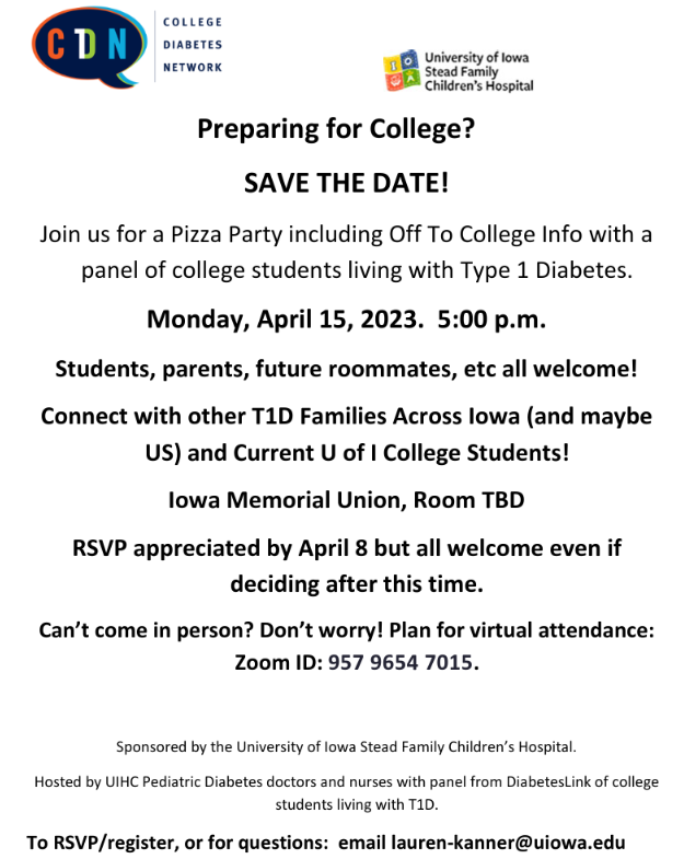
Type 1 Diabetes Prep for College Event to be Held April 15, 2024, 5 pm

Save The Date! Dr. Kanner has announced that she will host our annual College Preparation Event on April 15, 2024 at 5 pm. The event focuses on preparation for college success for those with type 1 diabetes. Students, parents, future roommates, and any others interested are welcomed to attend. If you are interested, contact us, either through the email link at Dr. Kanner’s webpage (link) or by contacting our diabetes nursing team. The event’s flyer is immediately below.
Grand Rounds : When Does Overnutrition Become an Endocrine Problem?

On February 16th, 2024, Drs. Kanner, Ramakrishna, and Parra Villasmil jointly delivered Pediatric Grand Rounds. Their talk was entitled “When Overnutrition Becomes an Endocrine Problem…Food for Thought“. They focused on the endocrine complications that can occur as a consequence of overweight and obesity in children and adolescents. Additionally, they touched on the practicalities of providing care to obese youth.
In the United States, nearly 1 in 5 children and adolescents are obese. In Iowa alone, over 50,000 youth are obese, per 2019 estimates (reference). Due to these high numbers, specialized obesity clinics are not a currently tenable solution. While most obese youth won’t develop endocrine complications, some may experience conditions such as type 2 diabetes, dyslipidemia, or polycystic ovarian syndrome.
Our Division accept referrals when these endocrine complications arise. Additionally, we welcome referrals for those rare cases where obesity is linked to endocrine conditions such as hypothyroidism or Cushing syndrome. Another reason for referral to endocrinology is when an obese adolescent and their family are prepared for the arduous process of bariatric surgery.
We extend our thanks to Drs. Kanner, Ramakrishna, and Parra Villasmil for sharing their expertise!
Gaining Better Understanding of Blood Sugars Problems Early in Cystic Fibrosis

Almost 10 years ago, investigators from our Division determined that young kids with cystic fibrosis (CF), less than 5 years of age, often have high blood sugars in response to a standardized sugary drink. However, the long term importance of these findings is unknown. Furthermore, we don’t know if this issue occurs when young kids are eating their usual foods and drink. To address this shortcoming, Dr. Katie Larson Ode of our Division, has partnered with other researchers across the country to create a study using wearable continuous glucose monitors. In one part of the study, they are using these monitors to determine what blood sugars do in young kids with CF in their usual environment (home, school, etc). However, so little is known about what blood sugars do in healthy young kids that it is difficult to know exactly what is normal. To address this, Dr. Larson Ode and her research partners will also study young, healthy kids. Dr. Larson Ode has just received grant funding to conduct the study, entitled “BEGIN Substudy: Continuous Glucose Monitoring in Healthy Children“. We thank Dr. Larson Ode and her research volunteers for their work to help advance knowledge.